
Hard x-ray bio imaging
Our research
X-ray imaging technologies are essential for life science, from basic research to clinical practice. Novel modalities such as, e.g., phase-contrast imaging [1], show promise for higher spatial resolution and improved contrast while still operating with an acceptable dose. Many imaging methods are source-limited.
We invented the liquid-metal-jet-anode hard x-ray source [2]. This compact electron-impact microfocus source potentially allows for more than 100x higher brightness than any competing laboratory x-ray tube. We exploit the unique high spatial coherence of the source for very-high-resolution propagation-based phase-contrast imaging at low dose [3]. Past projects include, e.g., microangiography in mouse, demonstrating detection of sub-10 µm diam vessels. The method was also extended to imaging of intact tumor microvasculature (cf. Fig. 1) [4] as well as to whole-body mouse CT. Current projects are:
Phase-contrast X-ray imaging
We develop laboratory X-ray phase-contrast imaging for samples where conventional absorption contrast does not show enough. Our liquid-metal-jet microfocus source combines high brightness with a small source spot, which gives the spatial coherence that propagation-based phase contrast needs — and with it soft-tissue contrast and near-cellular resolution in three dimensions, in an ordinary laboratory rather than at a synchrotron.
The method is non-destructive: the sample is imaged intact, with no sectioning and no staining, and can afterwards be processed further. That combination — 3D, unstained, micrometre-scale, laboratory-based — opens up applications that have little to do with each other. Two of them are shown below: tissue from cancer surgery, and a hand from a 2,500-year-old Egyptian mummy.
Clinical applications: 3D X-ray histology
Classical histology gives excellent microscopic detail, but it is two-dimensional, destructive and slow — typically several days from surgery to answer. When the question is whether a tumor has been fully removed, that delay matters: corrective surgery then means a second operation.
Together with pathologists and surgeons at Karolinska University Hospital and Karolinska Institutet, we have investigated whether phase-contrast X-ray microtomography can provide the same tissue-level information in 3D, and much faster. In a proof-of-concept study on twelve tumors — liver, pancreas and sarcomas — samples were fixed in a single acetone step instead of the usual multi-day formalin and alcohol series, imaged at around 10 µm resolution, and then compared with conventional H&E histology of the same samples. The two agreed well for the liver and pancreas tumors, and reasonably for the morphologically more difficult sarcomas.
Selected publications
J. Romell, C. Fernandez Moro, B. Brodin et al., “X-ray histology on rapidly fixed fresh tumor tissue samples for fast resection margin assessment”, Scientific Reports 16, 21642 (2026). doi.org/10.1038/s41598-026-61069-6
W. Twengström, C. F. Moro, J. Romell et al., “Can laboratory x-ray virtual histology provide intraoperative 3D tumor resection margin assessment?”, Journal of Medical Imaging 9, 031503 (2022). doi.org/10.1117/1.JMI.9.3.031503
Historical and archaeological applications
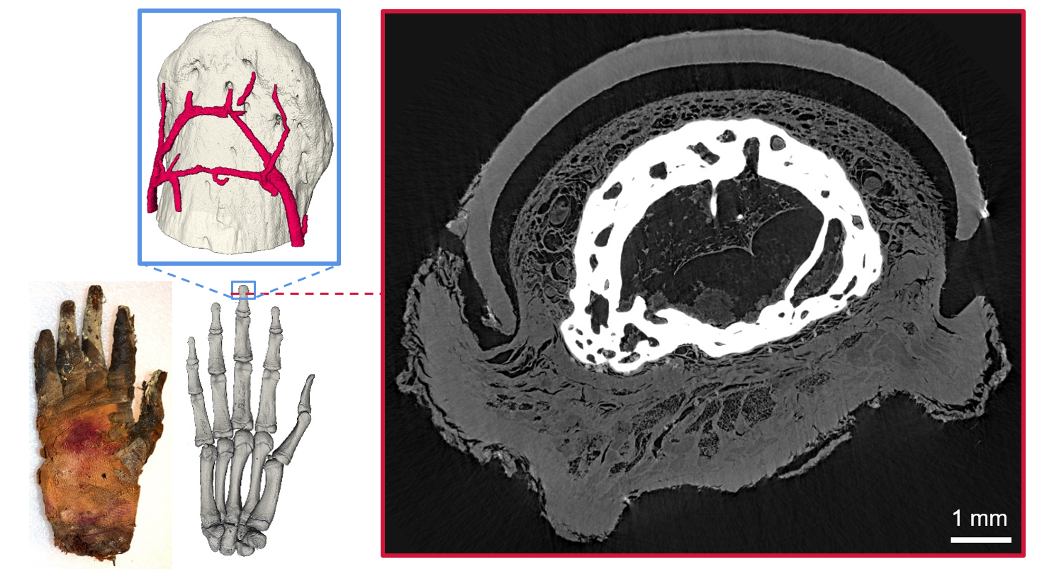
The same sensitivity to weakly absorbing material works on tissue that is thousands of years old. In collaboration with Egyptologists and museum researchers, we imaged a mummified human hand from ancient Egypt, roughly 2,500 years old, using propagation-based phase-contrast CT.
The whole hand was scanned, and a detailed scan of the fingertip reached an estimated resolution of 6–9 µm. Soft tissue turned out to be remarkably well preserved: tendons, nerves and blood vessels were visible, along with the separate layers of the skin, remnants of fat cells, and the vessels of the nail bed.
Nothing was cut, sampled or stained — which is the point. For museum and archaeological collections, where the specimen is irreplaceable, phase-contrast CT offers anatomical and paleopathological information at a scale that would otherwise require destroying part of the object.
Publication
J. Romell, W. Vågberg, M. Romell, S. Häggman, S. Ikram and H. M. Hertz, “Soft-Tissue Imaging in a Human Mummy: Propagation-based Phase-Contrast CT”, Radiology 289, 670–676 (2018). doi.org/10.1148/radiol.2018180945
X-ray fluorescence tomography
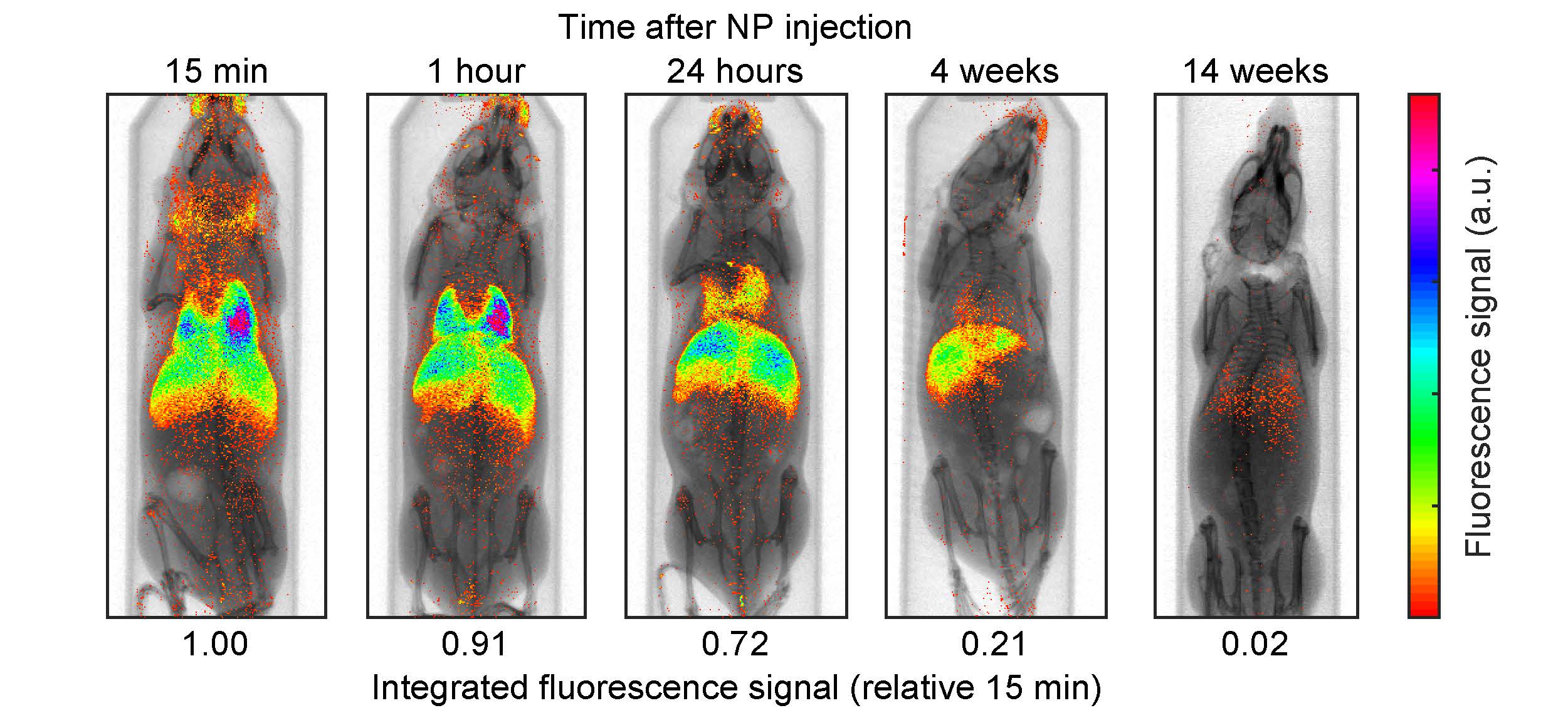
X-ray fluorescence (XRF) is used for 3D molecular imaging. We have developed a laboratory system for high-resolution imaging of small animals [10].
Work with us
For collaborations or student projects, please contact us.
References
- See, e.g., F. Pfeiffer et. al, Natute Phys 2, 258 (2006).
- O. Hemberg et al, Appl. Phys. Lett. 83, 1483 (2003).
- T. Tiuhimaa et al, Appl. Phys. Lett. 91, 074104 (2007).
- U. Lundstrom et al, Phys Med Biol. 59, 2801-11 (2014).
- W. Vågberg et al, Sci Rep. 5, 16625 (2015).
- W. Vågberg et al, Sci Rep. 8, 11014 (2018) .
- I. Häggmark et al, Opt. Express 25, 33543 (2017) .
- J. Romell et al, Radiology 289, 670-676 (2018) .
- I. Häggmark et al, IEEE TMI 40, 539-548 (2021) .
- K. Shaker et al, IEEE TMI 39, 3910-3919 (2020) .